Diatoms: Microphotographs by Paul Hargreaves and Faye Darling (17 photos)
The objects depicted in these astonishing photographs may appear alien, but they are actually magnified cells of living organisms—diatoms. Oceanographer Paul Hargreaves used an electron microscope to photograph these creatures, and artist Faye Darling retouched the images using specialized computer programs. 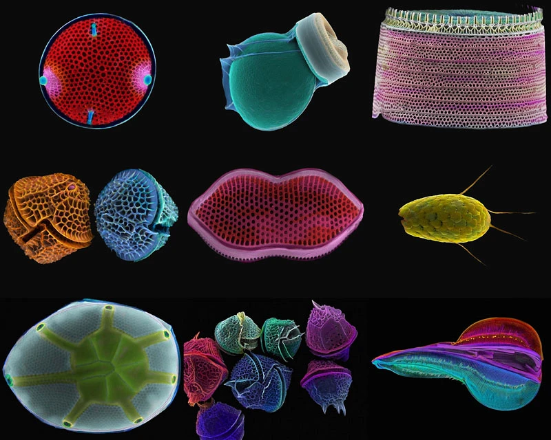
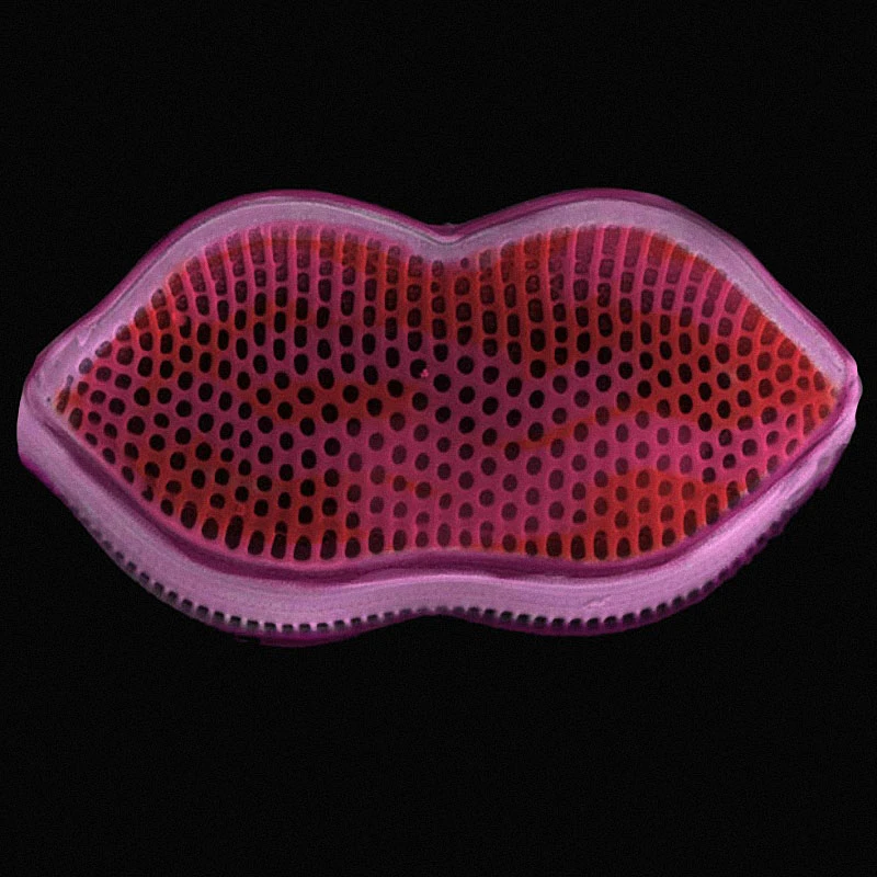
This photo may look like Salvador Dalí's "The Face of Mae West," but it's actually an electron microscope image of a species of diatom—a tiny, single-celled sea creature invisible to the naked eye. 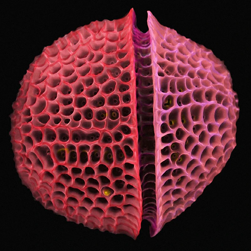

Как садят рис
Смотреть видеоThis is a photograph of a dinoflagellate—we see the dorsal part of the alga Protoceratium reticulatum.
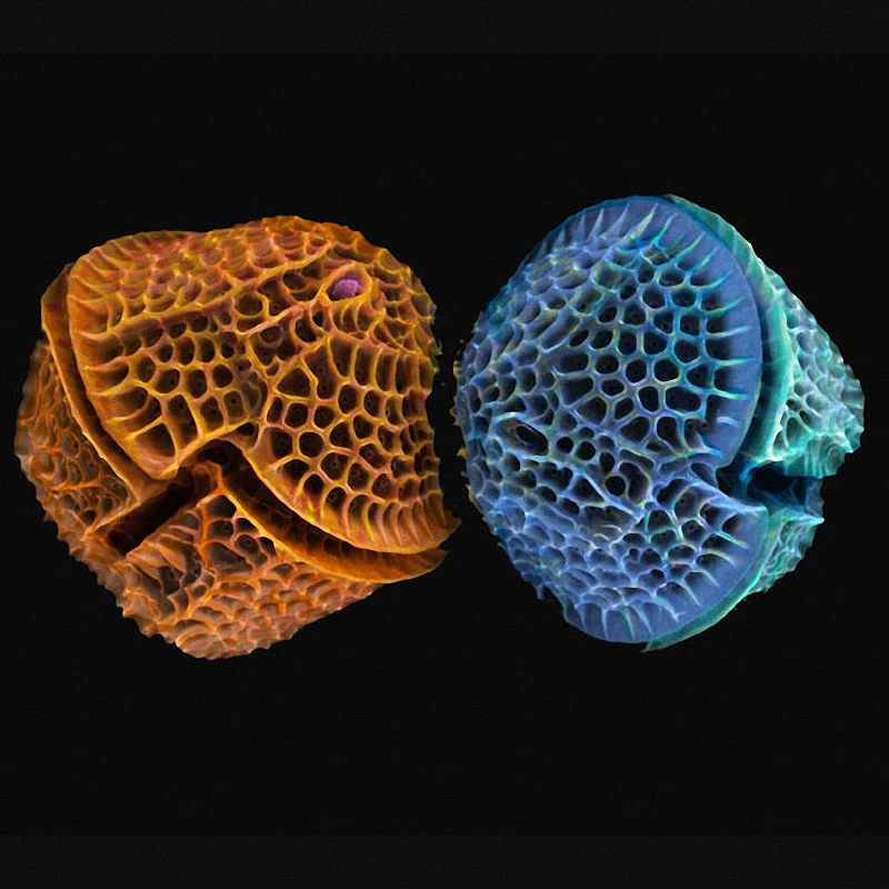
Dr. Paul Hargreaves and Faye Darling named this dinoflagellate "MiroMira2," although they look more like a pair of cupcakes.
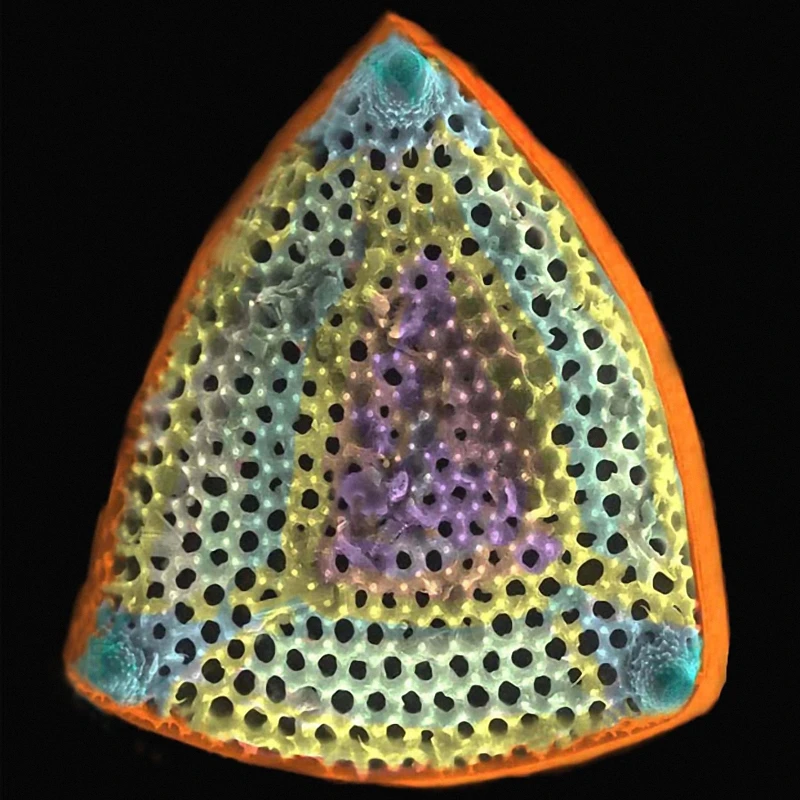
This creature looks like an iron steamer, although the authors called this photo "The Tibet Gem."
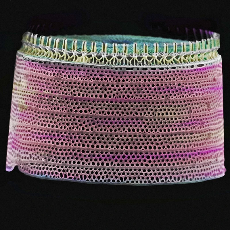
Here's another image, taken with an electron microscope and computer retouching, showing a diatom silicate alga.
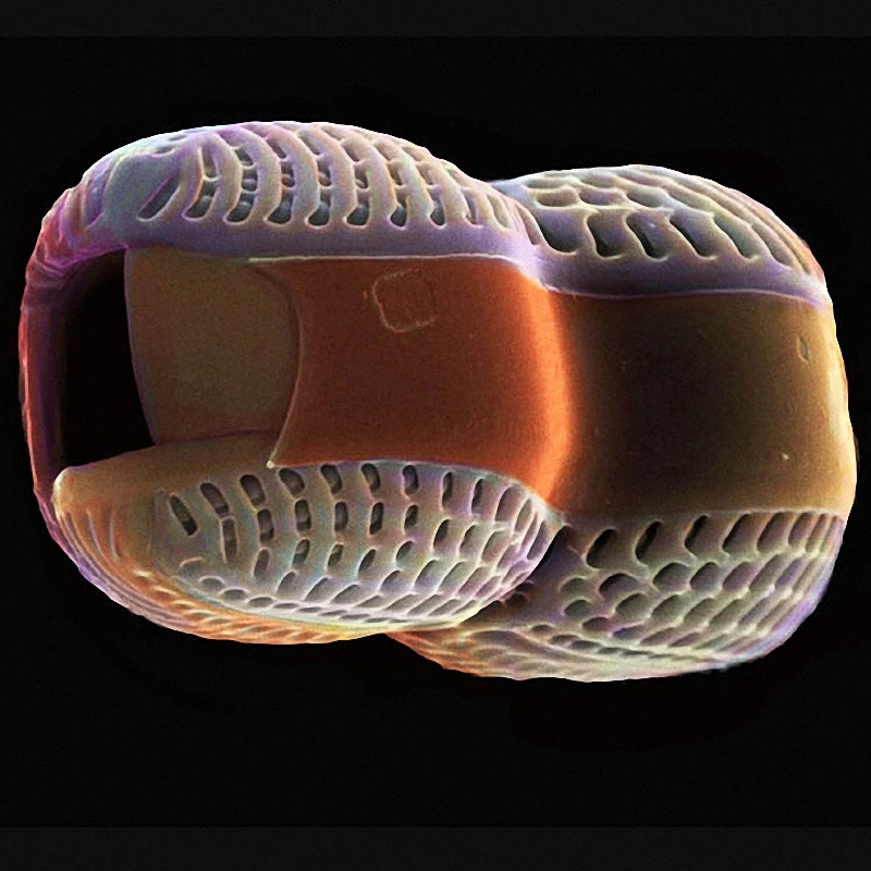
Dr. Paul Hargrivers and Faye Darling named this image "Peanut Opal."
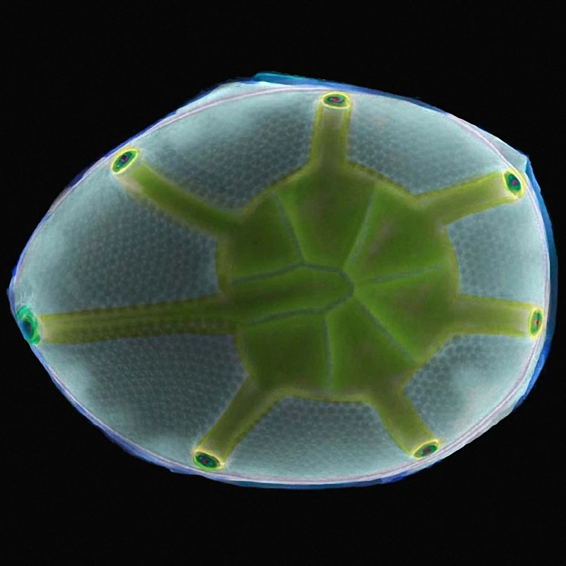
They dubbed this photograph "Blue Turtle."
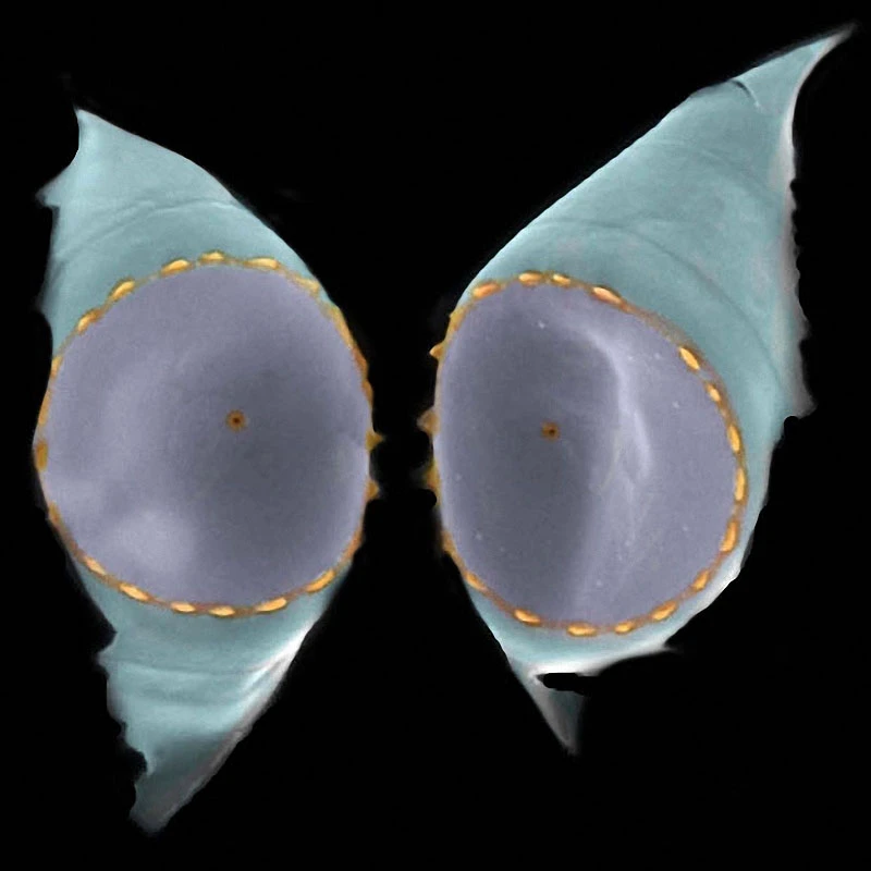
"Twin Crowns."
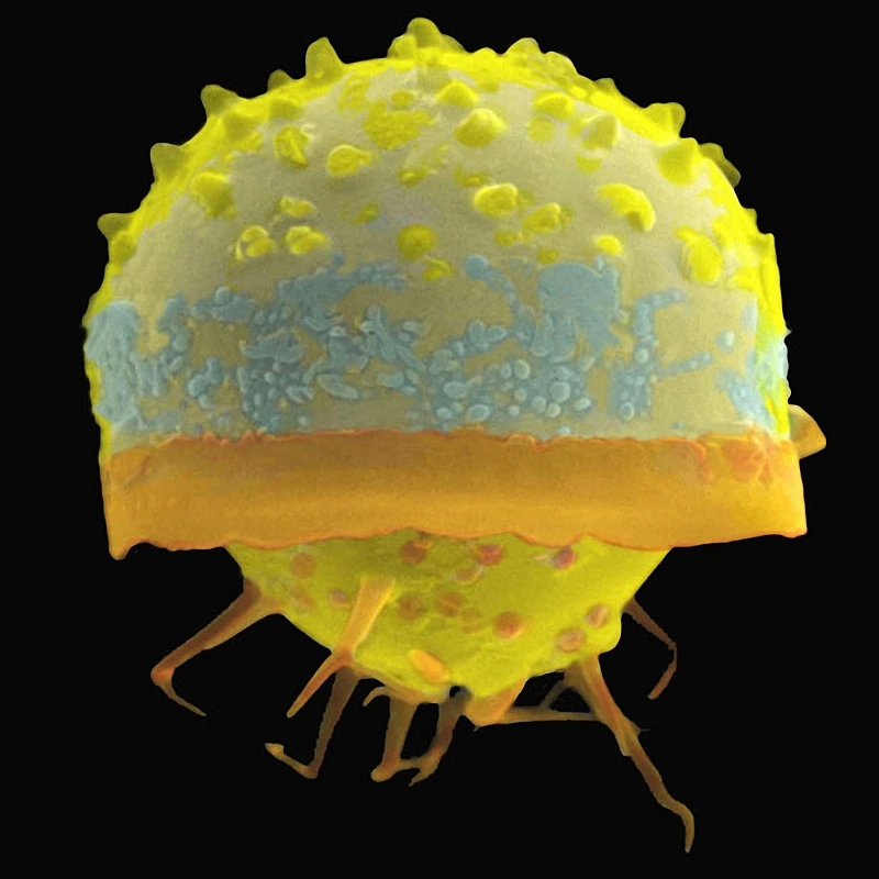
The authors dubbed this creature "Crooked Leg."
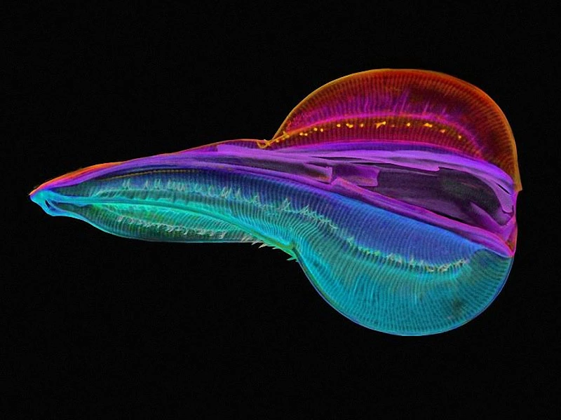
A specially colored cell of a diatom of the genus Entomoneis.
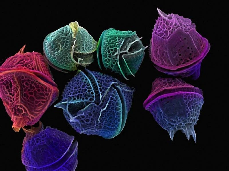
Images of dinoflagellates. Their population outbursts often lead to "red tides."
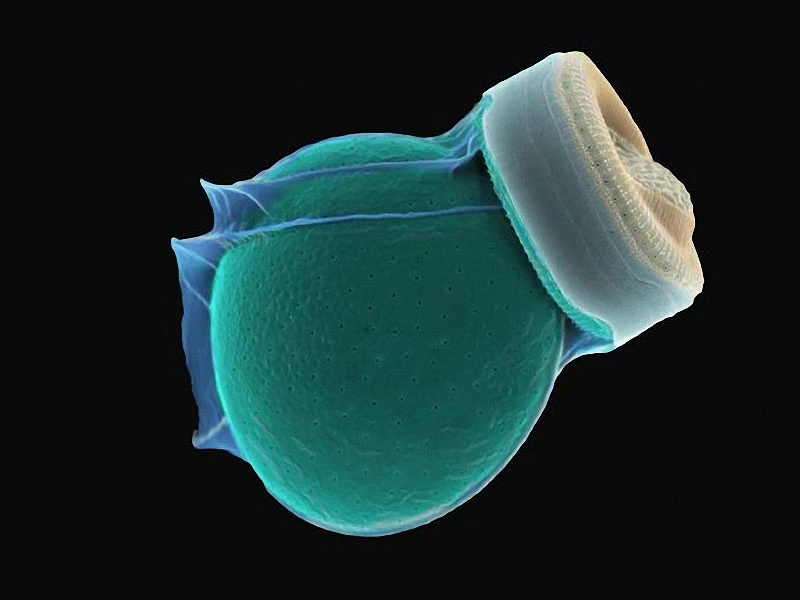
Structure of a fused dinoflagellate and a diatom.
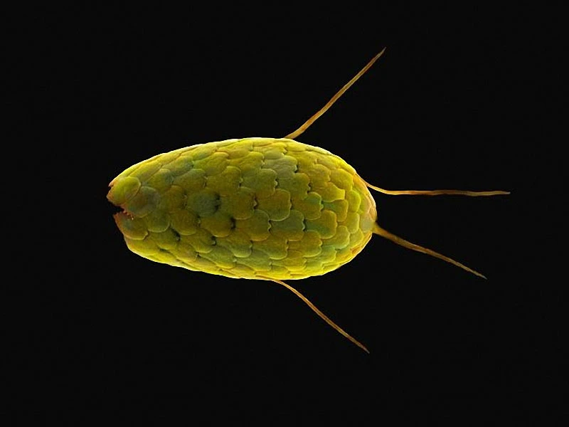
Amoeba.
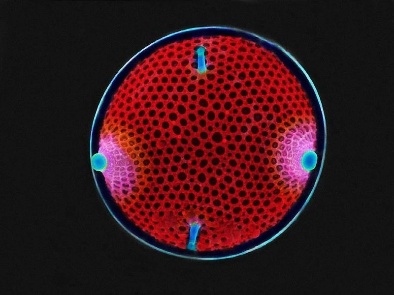
A structure called the "Wind Rose."
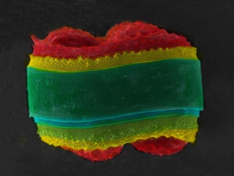
A diatom is a type of alga or phytoplankton that typically averages about 50.8 micrometers in size. These tiny creatures existed long before the dinosaurs.
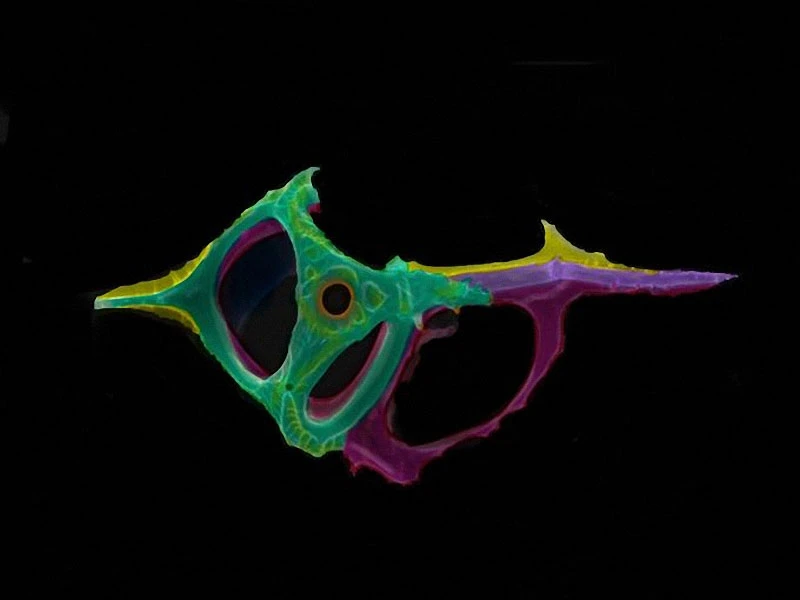
Ibridian.

Download
Archive size 2.86 Mb, downloaded 7 times