Incredible Photos of Tiny Creatures Under a Microscope (44 Photos)
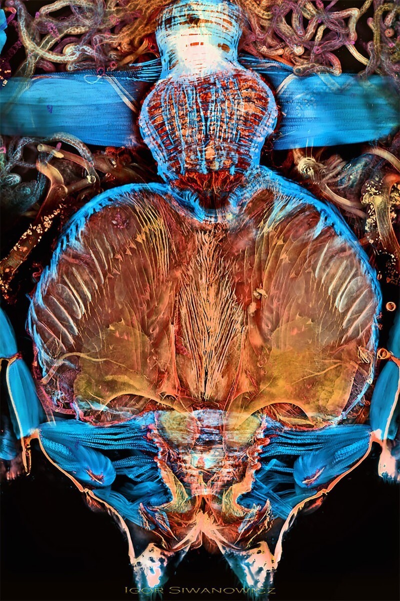
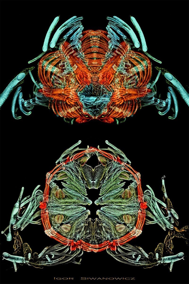
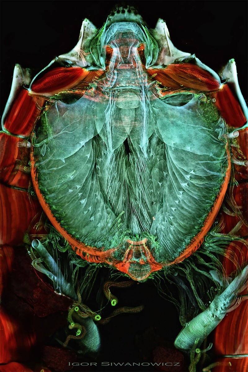
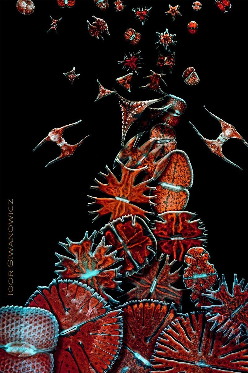
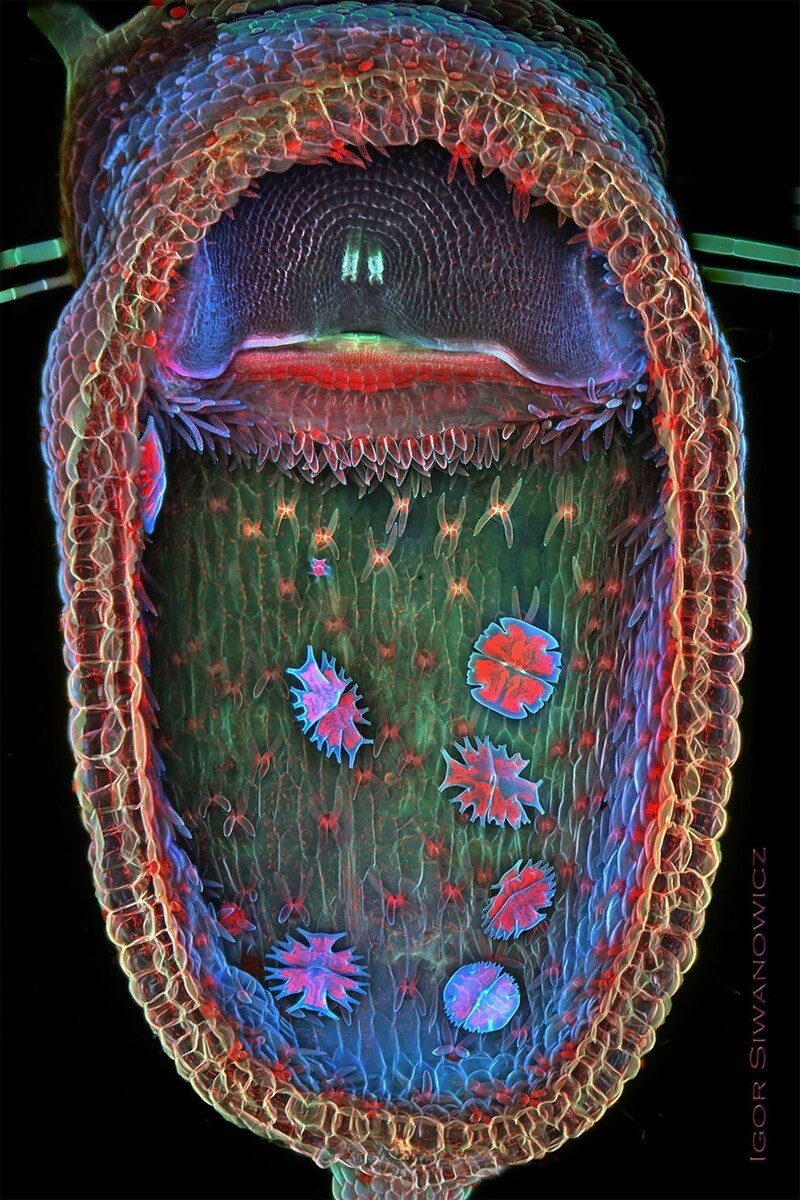
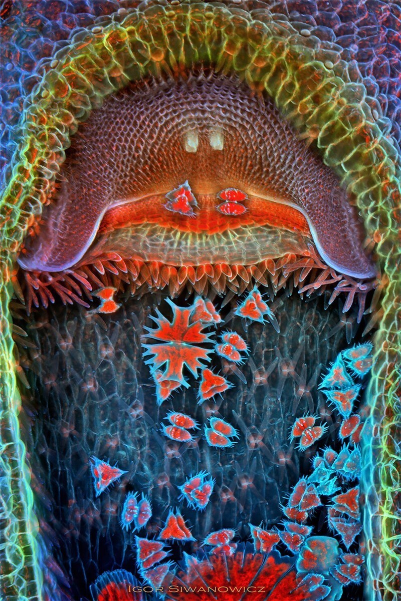
Photographer Igor Sivanovich showed us the wonders of life in kaleidoscopic color. This allows you to see in great detail the structure of the tiniest insects and plants.
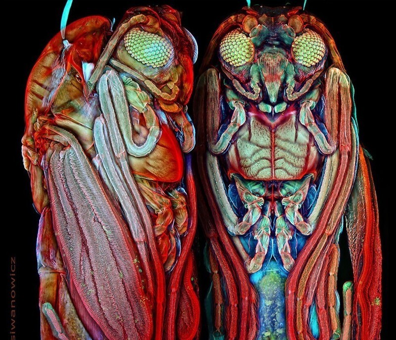

“A confocal laser scanning microscope takes photographs differently than a bright-field microscope (a microscope standard for biological research). This is a fluorescence microscope, which means that the sample being photographed is illuminated with light of one wavelength and at the same time emits light waves of another, longer wavelength. This is the same phenomenon that made the "invisible light" posters popular in the 1970s and 80s glow. A microscope that captures these rays takes a series of pictures of the small sample, scanning it point by point. Because the sample is much thicker than the plane of focus, a sequence of photographs—a series—is made by moving the sample up or down. From these “optical slices,” a three-dimensional image of the structures within the sample can be reconstructed,” he explains.


