30 impressive shots taken using a microscope (31 photos)
The winners of the 49th annual Nikon Small World Photomicrography competition have been announced. It involves photographers and scientists from all over the world, showing detailed photographs under a microscope. This year, about 1,900 works were submitted to the competition from photographers from 72 countries. First place was awarded to a photograph of the optic nerve head of a rodent. Let's take a look at these stunning photos that reveal many details that are invisible to us! 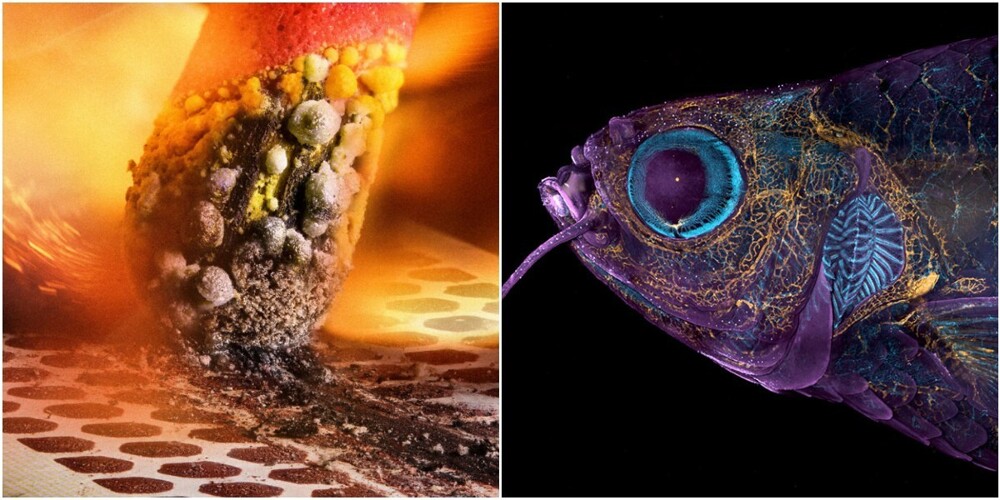
1. Sunflower pollen on an acupuncture needle. Photographer John-Oliver Dum 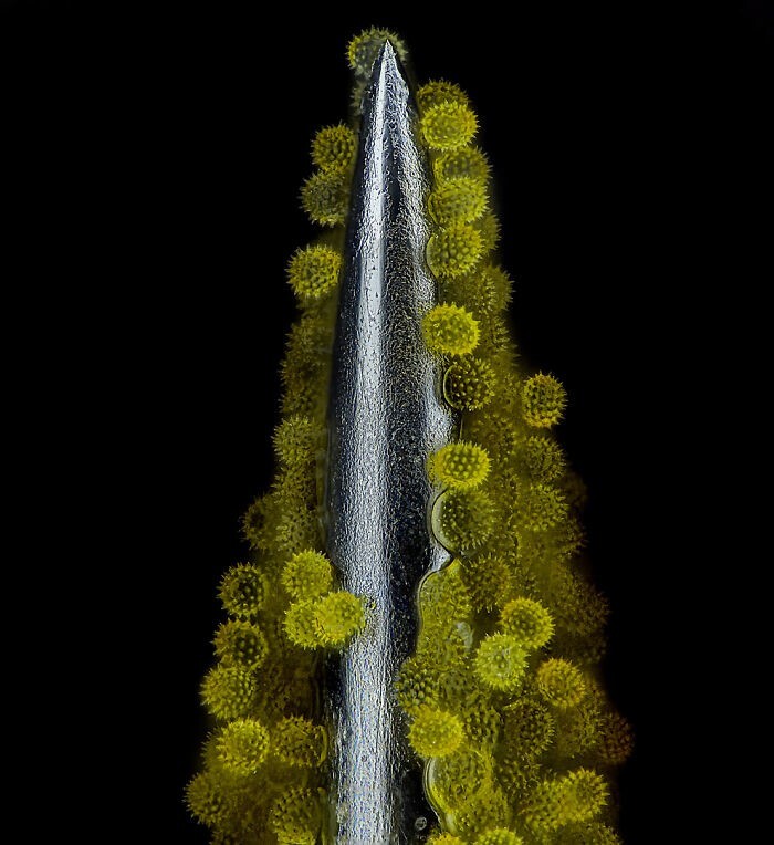
2. The moment of ignition of the match. The photo took second place. Photographer - Ole Bielfeldt 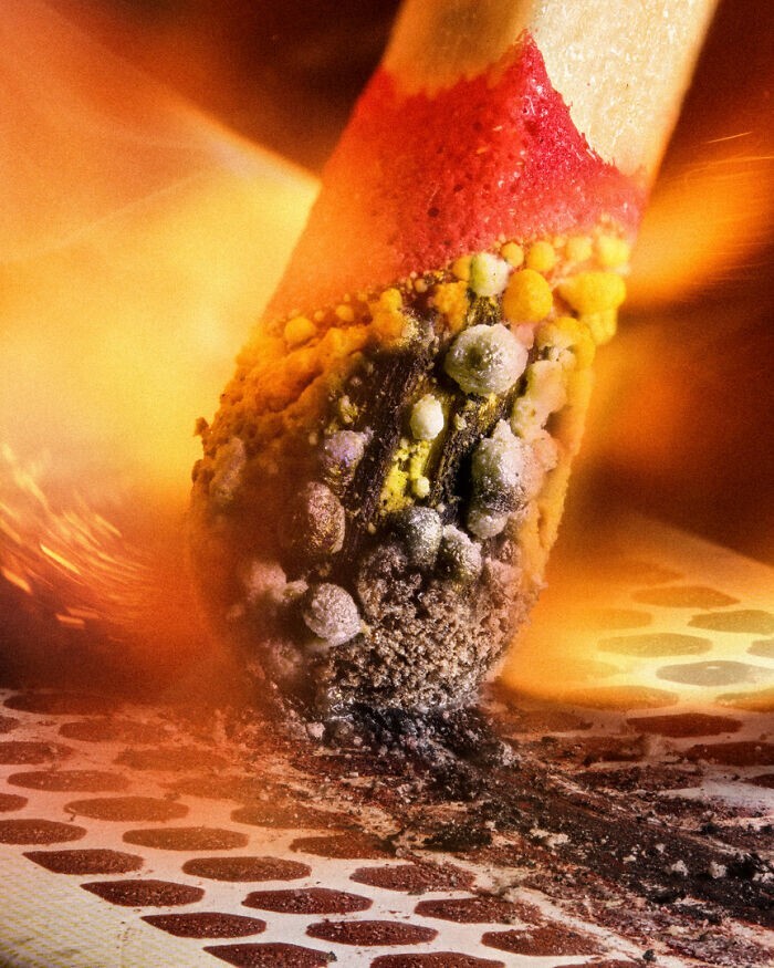

Как садят рис
Смотреть видео3. Caffeine crystals. Photographer Stefan Eberhard
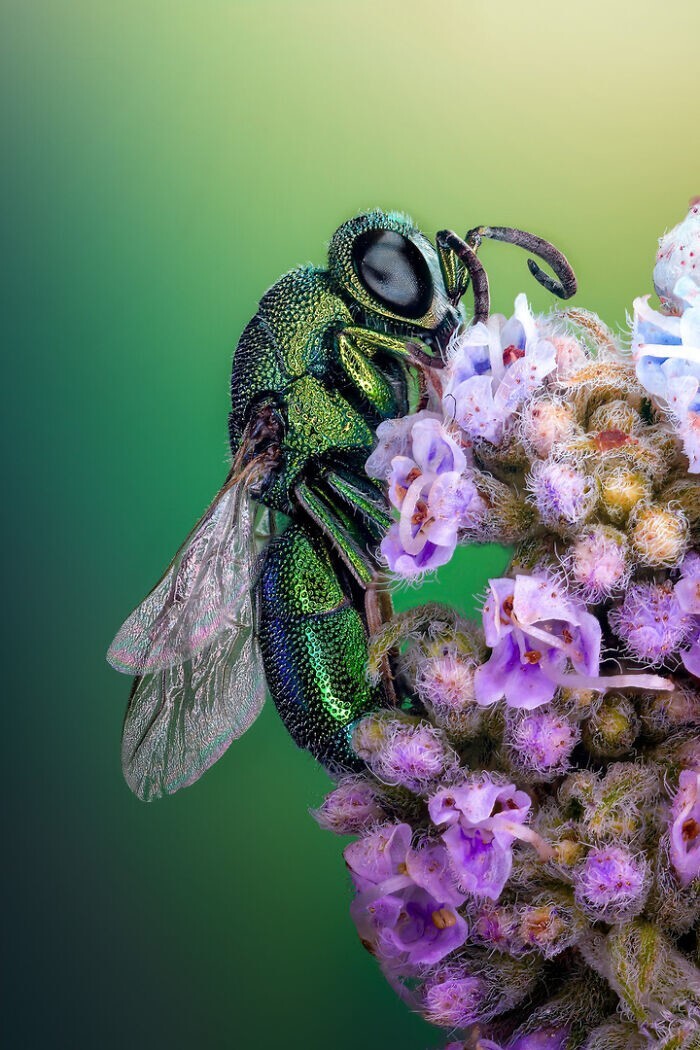
4. Spangled wasp on a flower. Photographer Sherif Abdallah Ahmed
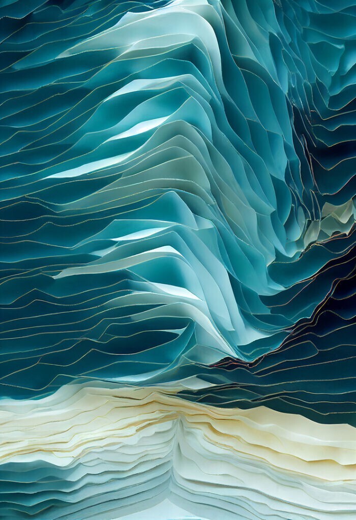
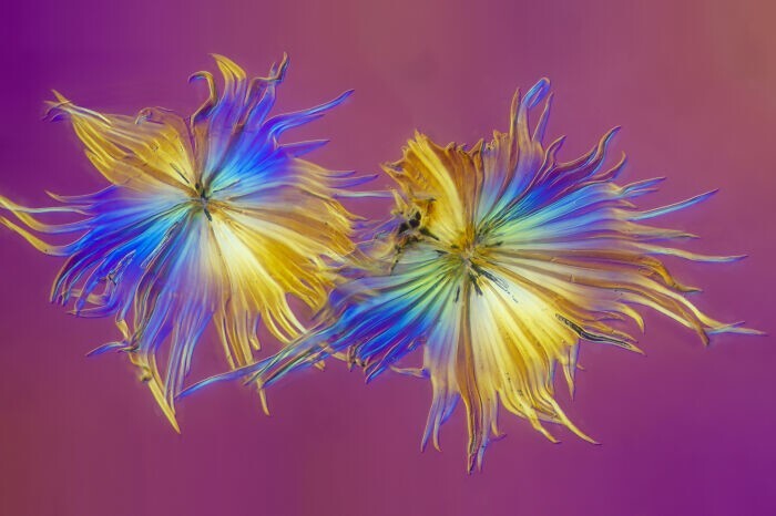
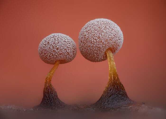
5. Crystallized sugar syrup. Photographer Dr. Diego Garcia
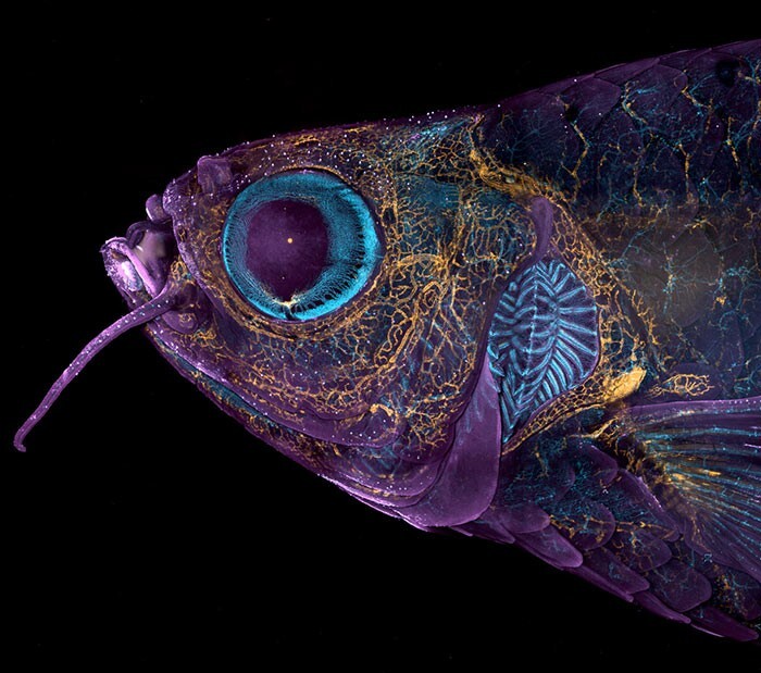
6. Fluorescent zebrafish, in which blood vessels (blue) and lymphatic vessels (yellow) are visible. Photographer Daniel Castranova
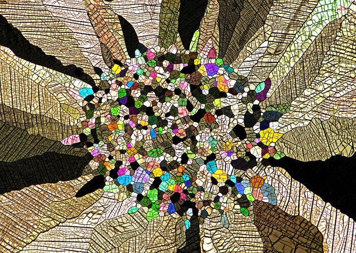
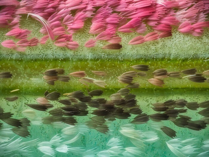
7. Wing scales of peacock butterflies Actias ningpoana under a microscope. Photographer Yuan Ji
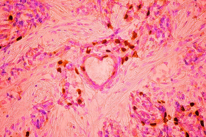
8. Breast cancer cells. The photo took third place. Photographer Malgorzata Lisowska
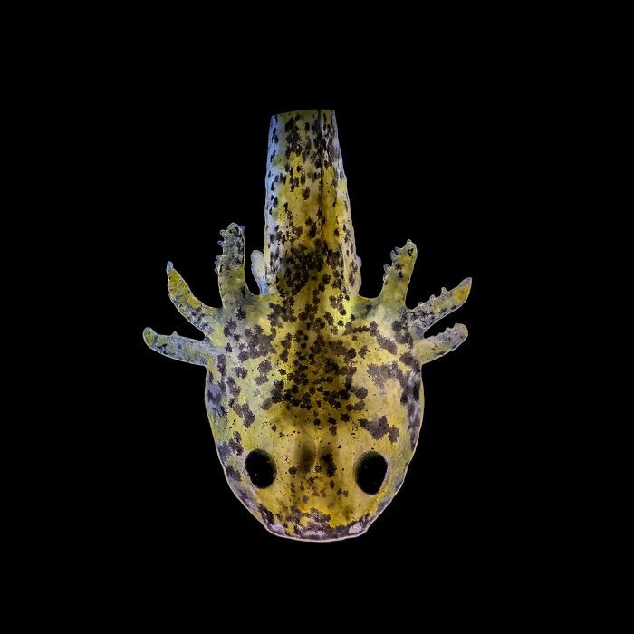
9. Axolotl (ambystoma larva), exactly one week old. Photographer Priscilla Vieto Bonilla
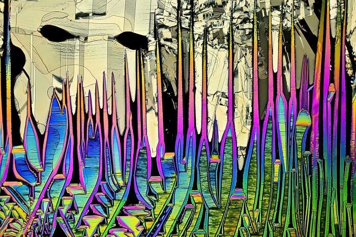
10. Malonic acid crystals dissolved in ethyl alcohol
11. Sea buckthorn trichomes are hairs that form pubescence on the underside of a sea buckthorn leaf. Photographer Walter Machielsen
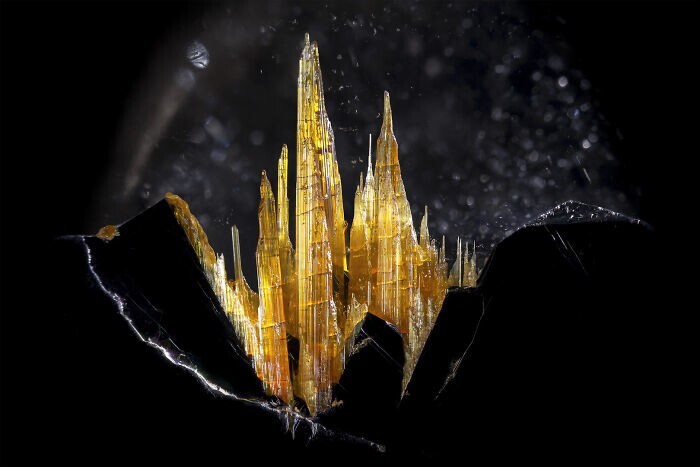
12. Gold rutile in quartz. Photographer Danny J. Sanchez
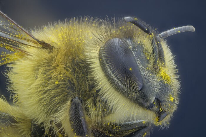
13. Bee. Photographer - Yusuf Ziya Özturk
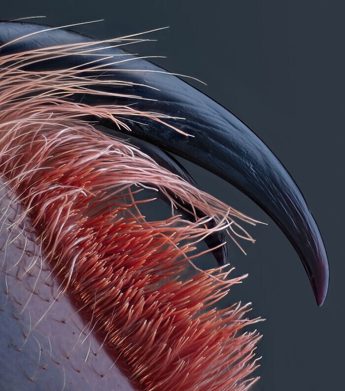
14. Poisonous fangs of a small tarantula. Photographer John-Oliver Dum
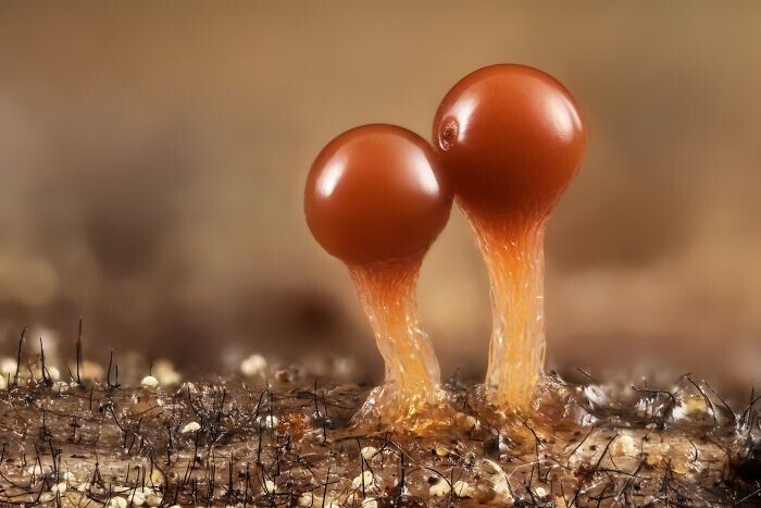
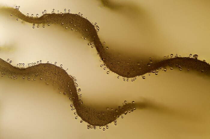
15. Slime mold (protozoa) Trichia crateriformis. Photographer Dr. Frantisek Bednar
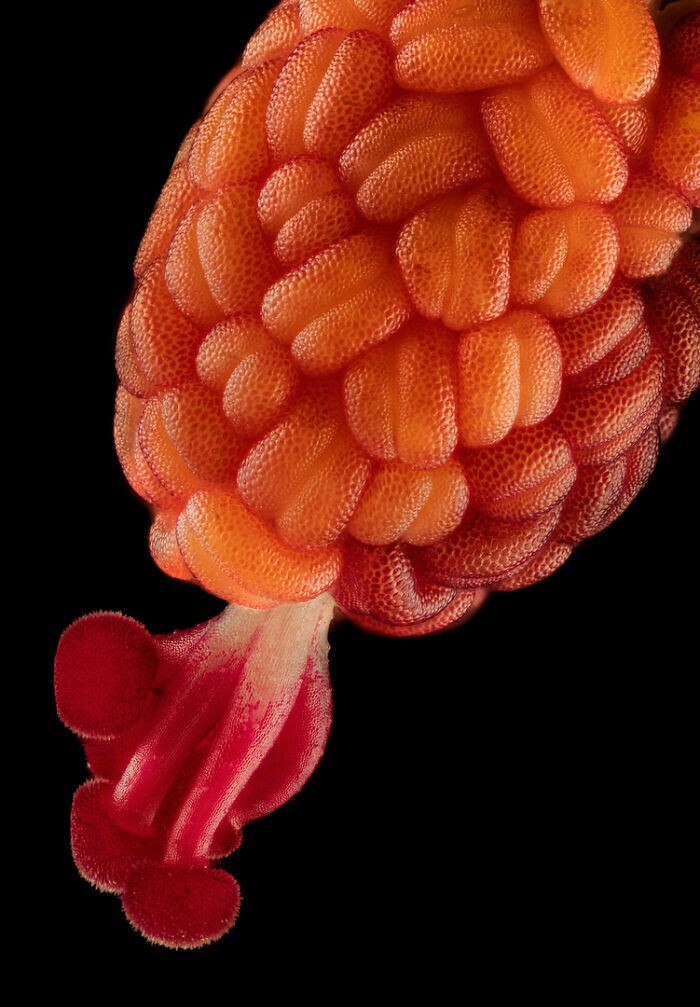
16. Developing stamen and stigma inside a hibiscus flower. Photographer Raghuram Annadana
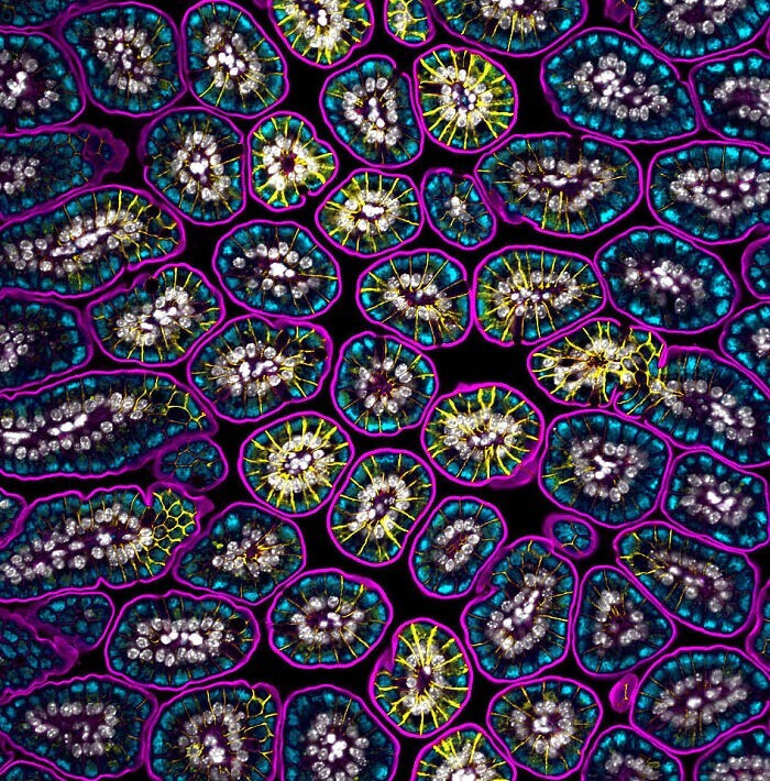
17. Cells of intestinal tissue of a newborn mouse. Photographer Dr. Amy Engevik
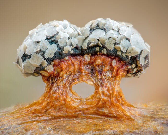
18. Slime mold Diderma tigrinum. Photographer Alison Pollack
19. Gills on the underside of the mushroom cap, on which sporophores are visible. Photographer - Charles B. Krebs
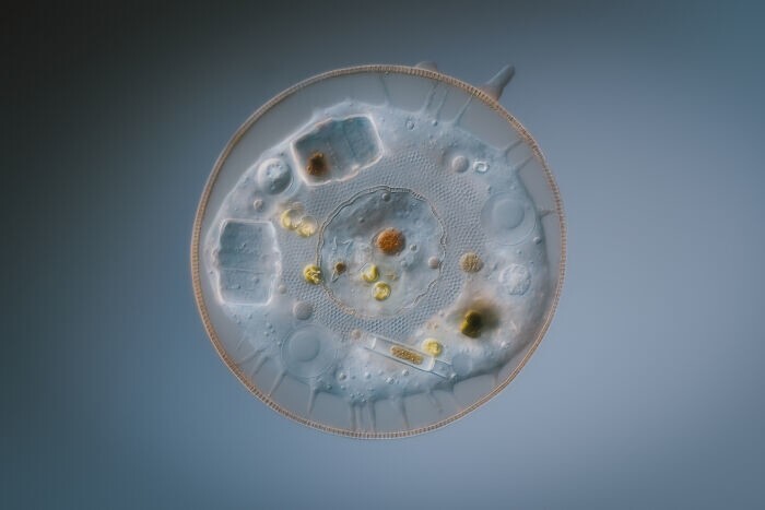
20. Amoeba. Photographer Dr. Håkan Kvarnström
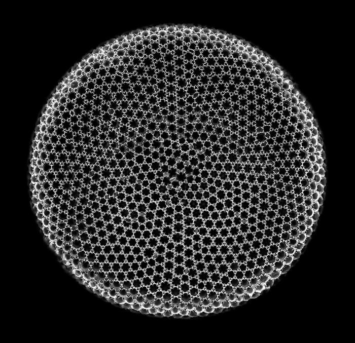
21. Fossil diatom. Photographer Michael Landgrebe
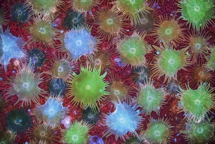
22. Coral (Acropora granulosa) fluoresces under blue light. Photographer Dr. Pichaya Lertvilai
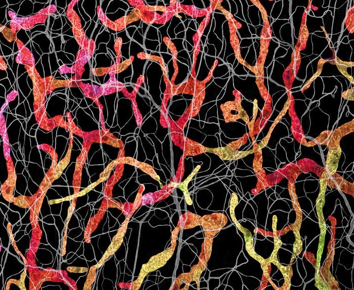
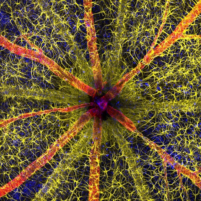
23. Rodent optic nerve head. Astrocyte cells (yellow), contractile proteins (red) and vascular cells are visible here.retinal shell (green). The photo took first place. Photographer - Hassanain Qambari
24. Slime mold Didymium sp. Photographer - Timothy Boomer
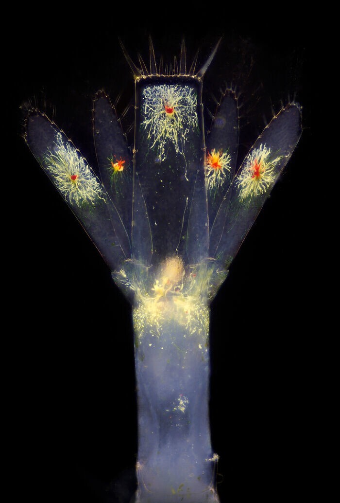
25. Tail of a planktonic shrimp larva. Photographer Ricardo Roberto Fernández Martinez
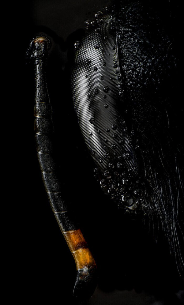
26. Head and antennae of a carpenter bee. Photographer Angel Navarro Gómez
27. Protective hairs covering the surface of leaves of Elaeagnus angustifolia, irradiated with ultraviolet light. Photographer Dr. David Maitland
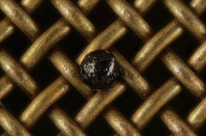
28. Cryptocrystalline micrometeorite on a laboratory sieve. Photographer Scott Peterson
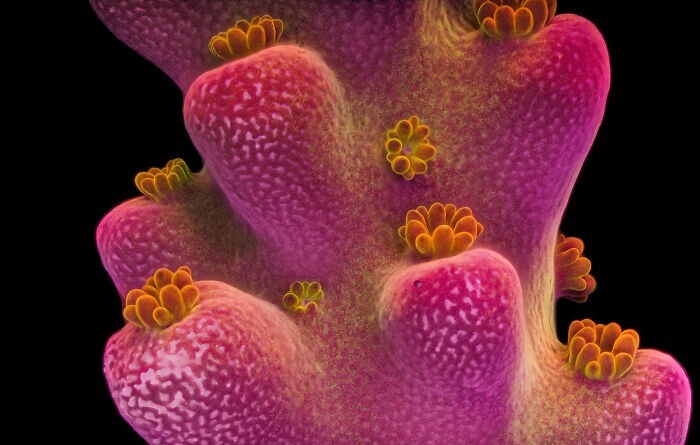
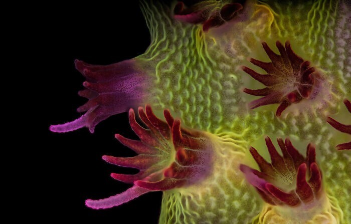
29. Acropora (polyp corals). Photographer - Dr. Pichaya Lertvilai
30. Blood and lymphatic vessels in the skin of the auricle of an adult mouse. Photographer - Satu Paavonsalo