The 25 best images from the Nikon Small World 2023 microphotography competition (26 photos)
The jury of the Nikon international photo competition selected the best photographs of this year taken using a microscope. The image of the optic nerve of a rodent was recognized as the best. The photograph by Hassanain Kambari and Jayden Dixon from the Australian medical research institute Lions Eye Institute not only has artistic value, but also contributes to the study of diabetic retinopathy. 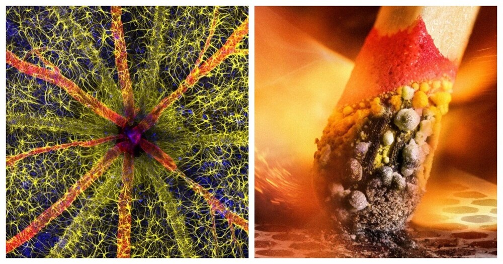
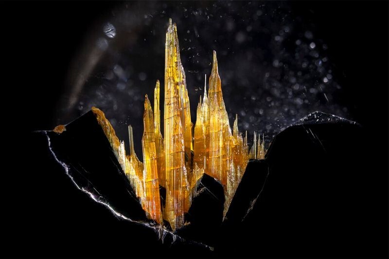
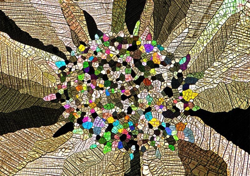
Rutile quartz is a type of quartz with needle-shaped inclusions.
Photographer: Danny Sanchez. 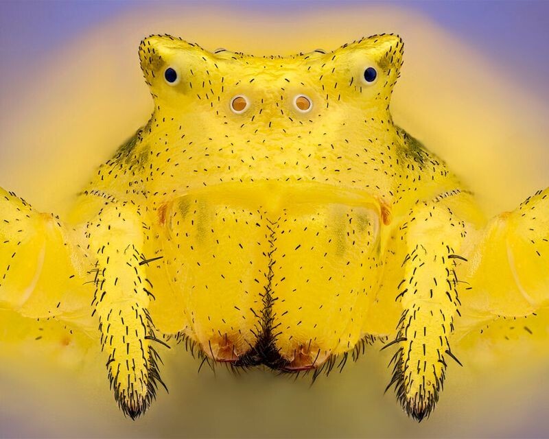

Как садят рис
Смотреть видеоCrab spider Thomisus onustus at 6.3x magnification.
Photographer: Sebastien Malot.
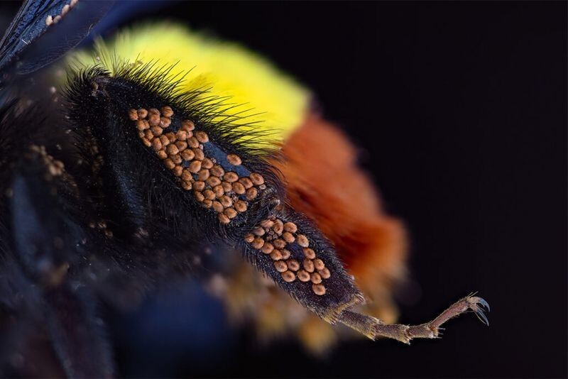
Phoretic mites on a bumblebee leg, photographed at 3x magnification.
Phoresia is the dispersal of an organism through its transfer to others.
Photographer: Amir Maqbool.
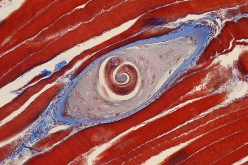
Trichinella in pork tissues.
Trichinella is a parasitic worm that causes trichinosis. Eating such meat can cause deadly complications.
Photographer: Dr. Nathan P. Myhrvold.
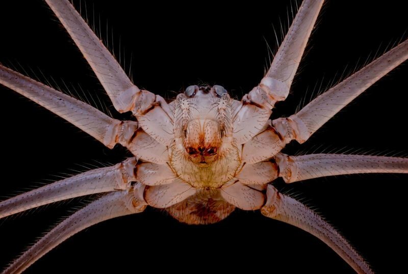
Underside of Pholcus phalangioides at 10x magnification.
Photographer: Dr. Andrew M. Posselt.
Top 20
Blood vessels (blue), lymph nodes (yellow), skin and scales (magenta) on the head of a transgenic zebrafish.
Photographers: Daniel Castranova and Dr. Brant M. Weinstein.
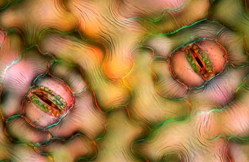
Stomata in a spathiphyllum leaf.
Photographer: Marek Mis.
Cryptocrystalline micrometeorite on a laboratory sieve.
Photographer: Scott Peterson.
Wing scales of the Actias ningpoana butterfly at 20x magnification.
Photographer: Yuan Zhi.
Carbon nanotubes photographed at 30x magnification.
Photographer: Dr. Diego Garcia.
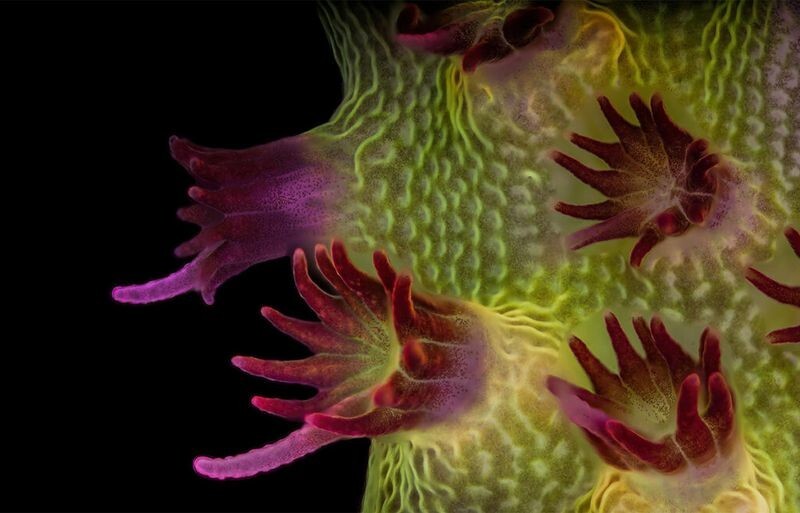
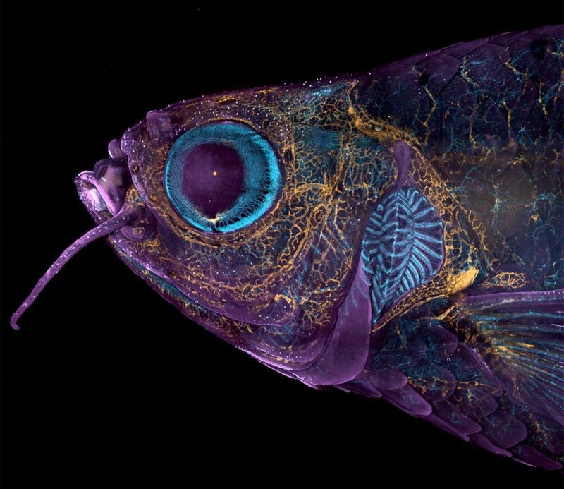
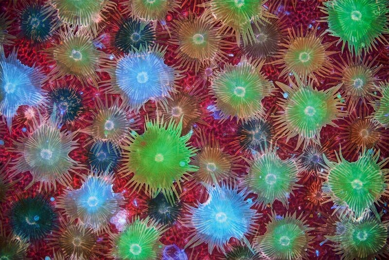
Fluorescent image of an Acropora coral showing individual polyps with symbiotic zooxanthellae.
Photographer: Dr. Pichaya Lertwilay.
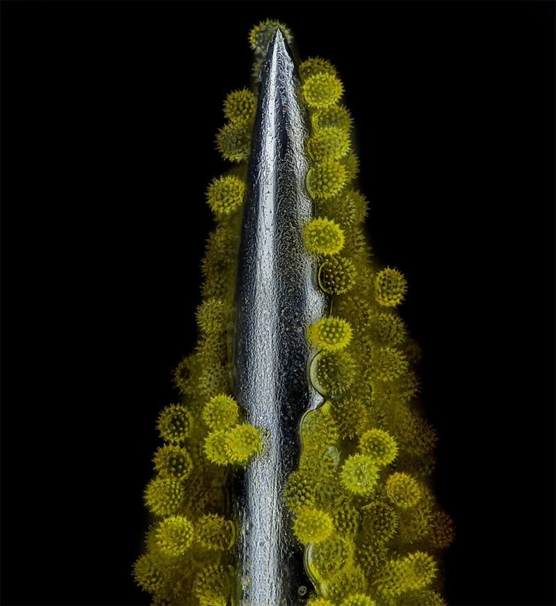
Sunflower pollen on an acupuncture needle, 40x magnification.
Photographer: John-Oliver Doom.
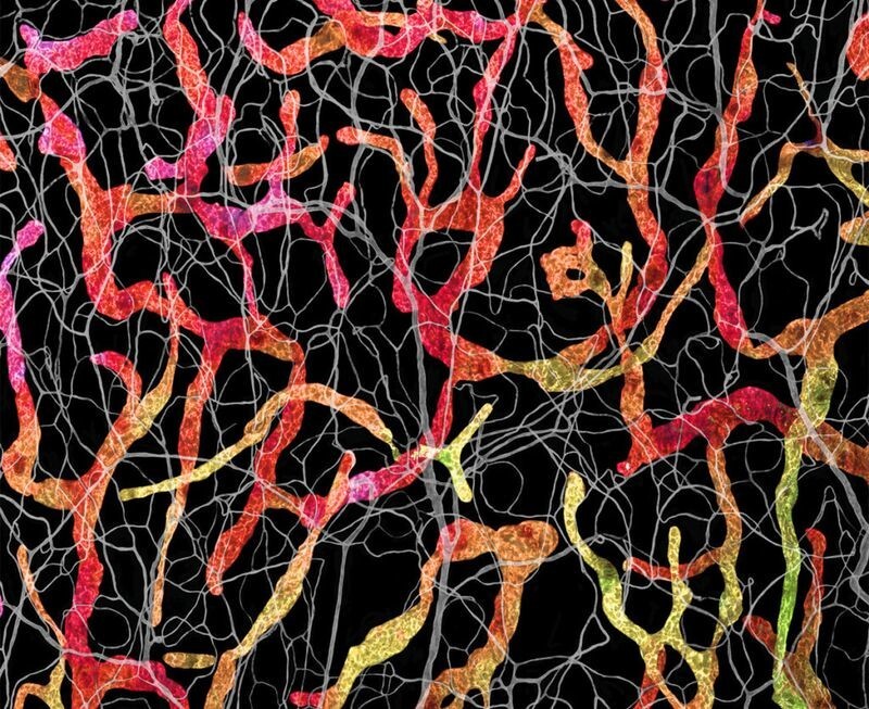
Blood and lymphatic vessels in the skin of a mouse ear.
Photographers: Satu Paavonsalo and Dr. Sinem Karaman.
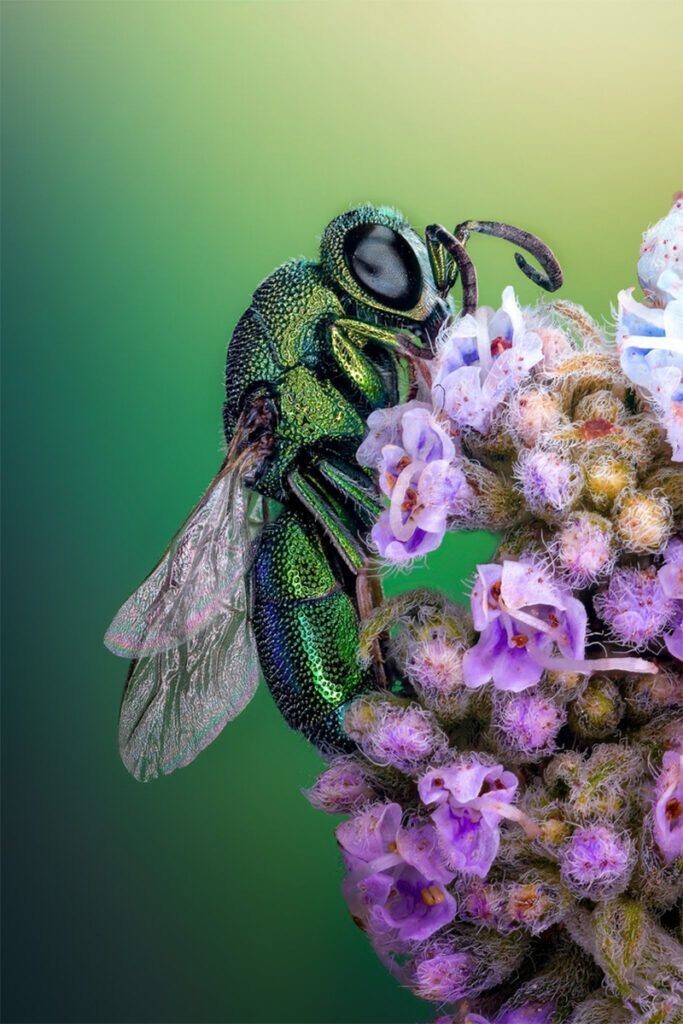
A glossy wasp on a flower in Tanta, Egypt.
Photographer Sherif Abdallah Ahmed.
Crystallized sugar syrup at 25x magnification.
Photographer: Dr. Diego Garcia.
Top 10
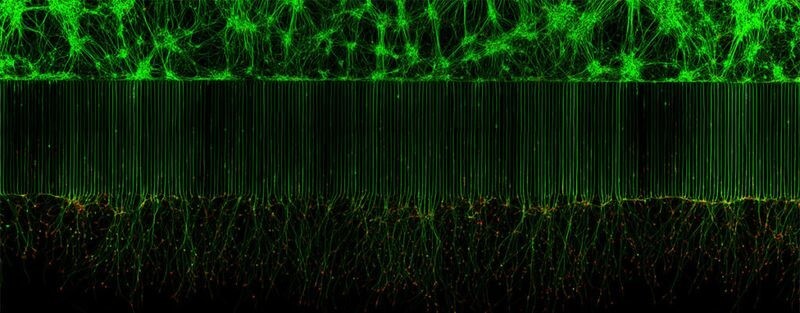
Motor neurons in a microfluidic device for separating cell bodies (top) and axons (bottom).
Photographers: Melinda Beccari and Dr. Don W. Cleveland.
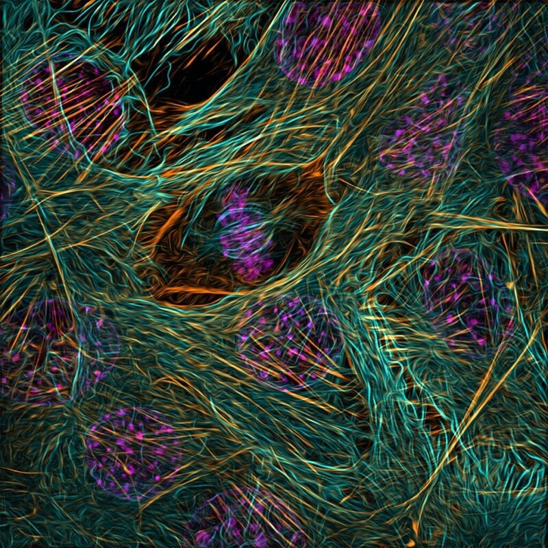
Cytoskeleton of a dividing myoblast.
Photographer: Vaibhav Deshmukh.
Caffeine crystals.
Photographer: Stefan Eberhard.
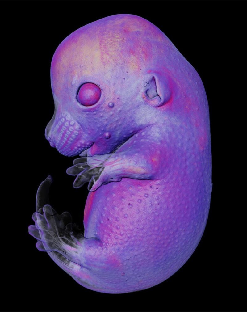
Mouse embryo at 4-cratomic magnification.
Photographers: doctors Grigory Timin and Michelle Milinkovic.
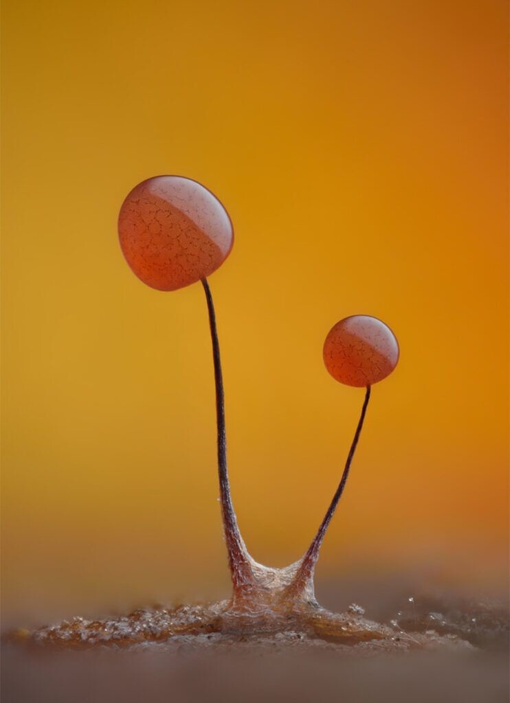
Slime mold Comatricha nigra, with capillary fibers visible through the translucent peridium.
Photographer: Timothy Boomer.
Autofluorescent protective hairs on the surface of leaves of Elaeagnus angustifolia.
Photographer: David Maitland.
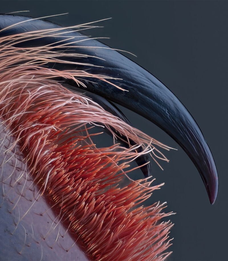
Poisonous fangs of a tarantula at 10x magnification.
Photographer: John-Oliver Doom.
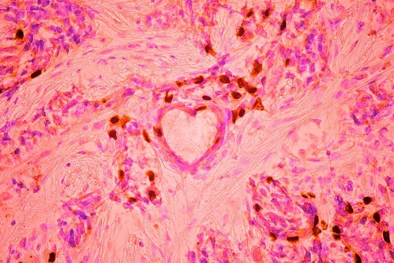
3rd place. Breast cancer cells at 40x magnification.
Photographer: Małgorzata Lisowska.
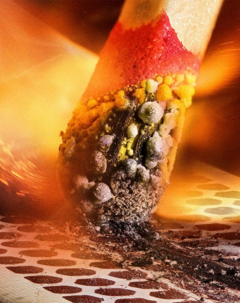
2nd place. Ignition of a match.
Photographer: Ole Bielfeldt.
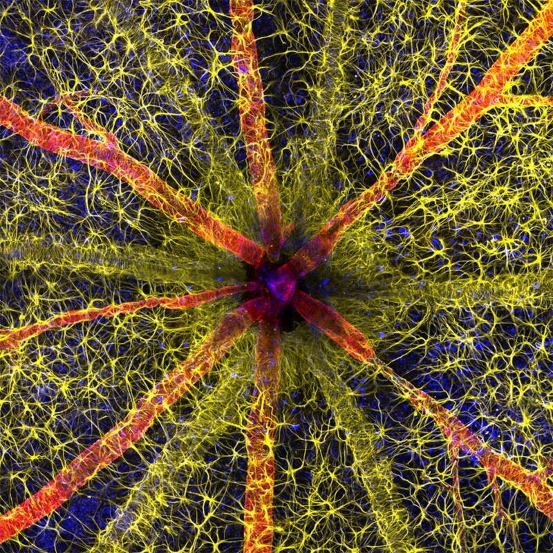
1st place. The rodent optic disc shows astrocytes (in yellow), contractile proteins (in red) and the retinal vasculature (in green).
Photographers: Hassanain Kambari and Jayden Dixon.
Diabetic retinopathy is damage to the retina of the eye. Every fifth diabetic patient worldwide suffers from this disease. Hassanain Kambari has been working for several years on methods for early diagnosis of the disease and reversing it.